
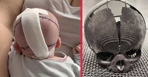
A baby girl born missing a portion of her skull had life-saving ‘unique’ surgery using 3D printing technology.
The child from Rzeszow, Poland, was born with a congenital abnormality that caused about one-fifth of her skull to not fully develop at the rear of her head.
The abnormality was missed during prenatal screenings and was discovered only after she was born in February.
Doctors were given four days to do surgery.
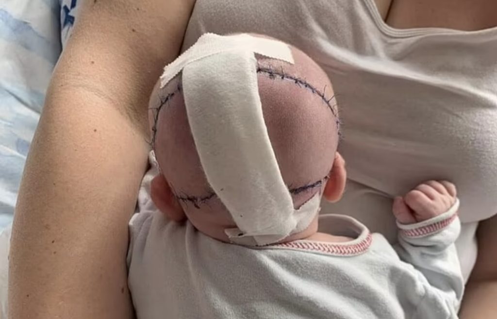
In a very sensitive two-hour process, they were able to patch her up using skin and soft tissue from different sections of her body.
The procedure went off without a hitch after doctors envisioned and practised on an identical copy of the kid’s skull.
They took comprehensive images of her skull and sent it to a 3D printing business, which created a 1:1 replica.
In Poland, doctors were able to practise fixing a newborn baby girl’s shattered skull with an exact 3D-printed copy, saving her life.
It exposed brain tissue, making her vulnerable to diseases that would very surely have killed her if left untreated.
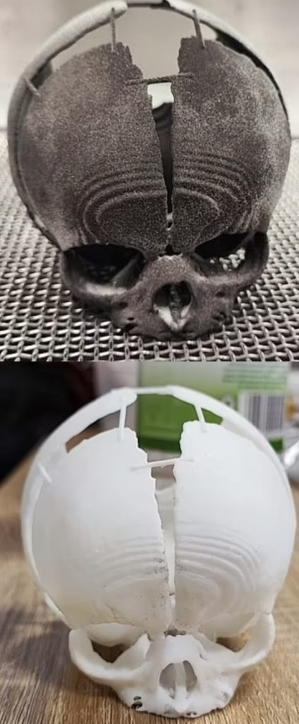
3D technology experts Sygnis created a three-dimensional prototype to assist surgeons in seeing the region of lost bone in real time. The researchers employed two separate technologies to ‘prevent disaster and offer surgeons with the most feasible options.’ Left: The first skull replica was created using selective laser sintering (SLS), a method that requires fusing nylon powder layers together. Right: For the second model, they also employed stereolithography (SLA), a technique in which photosensitive resins are hardened layer by layer.
WHAT EXACTLY IS AN ENCEPHALOCELE?
Encephaloceles are unusual birth malformations linked with skull deformities lead to incomplete bone fusion, resulting in a space through which a piece of the brain protrudes (protrudes).
Cerebrospinal fluid or the membranes that surround the brain (meninges) may also protrude through this space in rare situations.
The region of the brain that protrudes from the skull is normally covered by skin or a thin membrane, giving the defect the appearance of a tiny sac.
Protruding tissue can occur anywhere on the head, however it most commonly affects the rear of the skull (occipital area).
The majority of encephaloceles are severe and serious birth abnormalities that are detected before delivery.
In exceedingly rare circumstances, certain encephaloceles may be tiny and go undiscovered.
The specific aetiology of encephaloceles is unclear, however the illness is most likely the consequence of a mix of various causes.
In the UK, incidences are infrequent, with around 1.7 in per 10,000 births.
In most situations, encephalocele therapy consists of surgery to reposition the section of the brain that is outside the skull and seal the opening.
Neurosurgeons can frequently repair even big encephaloceles without harming the kid to lose any additional function.
This enables doctors to observe for themselves the degree of the bone loss and assisted them design the procedure.
Because the kid’s bones are still growing, doctors are holding off on repairing the missing portion of her skull.
In February, the daughter was born in a hospital in Rzeszow, in Poland’s south-east.
She was subsequently moved to Krakow, a specialty children’s hospital 100 kilometres away.
Krakow experts used CT and MRI images to create an accurate virtual reconstruction of her skull.
They transferred the model to a computer and shipped it to the country’s capital, Warsaw, for 3D printing with nylon and resins.
From beginning to end, the printing took 26 hours, with two skulls created at the same time and shipped back to doctors at the University Children’s Hospital.
The surgeons utilised the skulls to recreate the difficult process and identify any complications that may arise during surgery.
To avoid infection in her brain, the newborn was kept separate in an incubator. Through a tube, she was fed her mother’s milk.
Surgeons started the two-hour process of repairing the soft tissue of the head after examining the skulls and documenting the exact form and size of the defect.
Skin, muscle, and fat from other regions of her body were used in the procedure.
Professor Łukasz Krakowczyk, who performed the surgery, stated, ‘This is a very unusual problem, and this is the first time I have had to confront such a treatment in my 20 years of expertise.’
‘So that was a highly novel technique for me.’ A fifth of the skull surface was missing, indicating a significant flaw.
‘The procedure had to be done quickly since a portion of the brain was uncovered, threatening to contaminate the central nervous system.’
‘The therapy was planned using a model made using 3D printing technology.
‘This enabled for an accurate calculation of the bone loss, which considerably improved the operation’s design and scope, significantly decreasing the operation’s duration.’
The successful surgery occurred on February 28 but was just recently announced. She will now require more therapy as her skull bones develop.
‘The youngster is waiting for another procedure, this time the rebuilding of the skull bone, however we know that the bones are developing and that is why we need to wait for this stage of the operation,’ said Professor Karkowczyk.
‘3D printing will also be essential at the stage of skull bone defect restoration, when it will be necessary to correctly fit and design bone reconstruction.’
‘3D printing will also be crucial at the point of repair of the skull bone defect, when it will be important to accurately fit and plan bone rebuilding,’ said Professor ukasz Krakowczyk, who performed the procedure.
When the girl was born, her occipital bone, which forms the rear and base of the skull, was not fully grown.
In a race against the clock, the 3D-printing team employed two alternative technologies to ‘prevent failure and equip doctors with the broadest conceivable choices.’
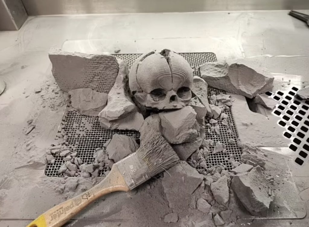
The first skull copy was created using selective laser sintering (SLS), a method that requires fusing nylon powder layers together.
For the second model, they also employed stereolithography (SLA), a technique in which photosensitive resins are hardened layer by layer.
‘The girl who was going to arrive in the world at any time did not have a formed occipital bone, which meant that the brain tissue was partially exposed,’ stated a Sygnis spokeswoman.
‘In the case of anatomical models, both SLS and SLA technologies are distinguished by excellent attention to detail.’
The models were developed in record time to help surgeons “anticipate the circumstances they would confront during the kid’s surgery.”
The SLS model was precise to 0.125mm detail and produced in a ‘relatively fast printing period,’ according to the business.
‘After the printing process was done, the print in SLS technology underwent a thorough cleaning of unbaked powder and sandblasting,’ stated Sygnis.
‘We completed the entire project in 26 hours.’